
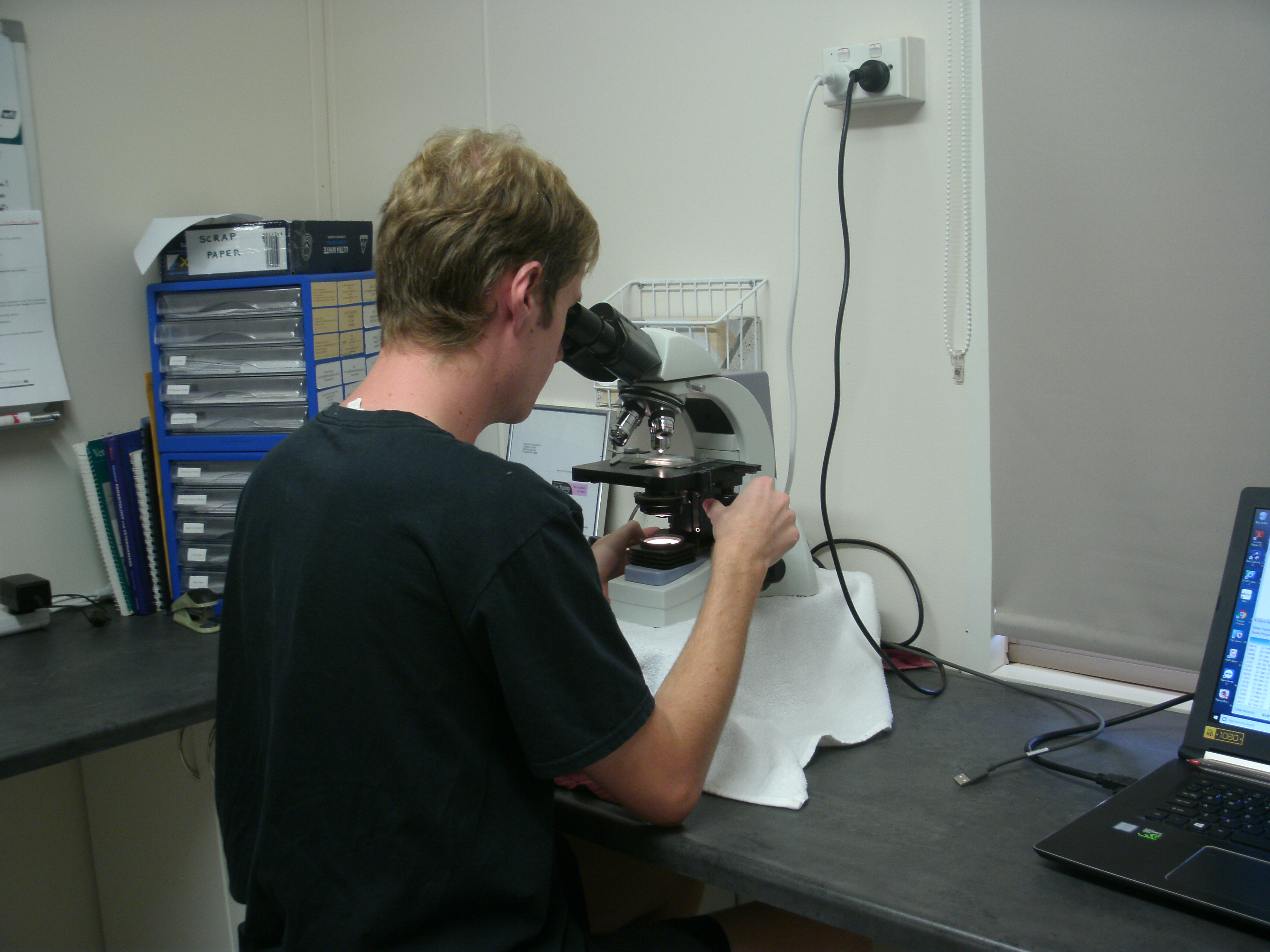
On a farm half an hour from Temora, in the middle of New South Wales, is a lab. Every day before work, Tom Shuttleworth drives to town, collects the mail, and takes it back to be analysed under a microscope. However, this isn’t anything that you’d normally find in a parcel. Instead, Tom looks at sheep poo.
You do what?!
I’ve been working with Tom for the past couple of years. I generally describe my job as ‘looking at sheep poo under a microscope’. We do this to check how many parasites are in the digestive systems of farm animals.
The more eggs we count, the more parasites there are, and the sicker the animal is. Farmers can use this information to treat their animals properly.
“Get it wrong with parasites and it can be a devastating blow,” says Tom. “Get it right and production levels [on the farm] can stay high.”
What is a parasite?
A parasite is an organism that lives on another (known as a host). Parasites get their nutrients from the host, often causing damage in the process.
Examining the excrement
Most of our samples are sheep poo, but we also test cows, horses and goats. These animals mostly eat grass or grains, so the poo just looks like really muddy grass.
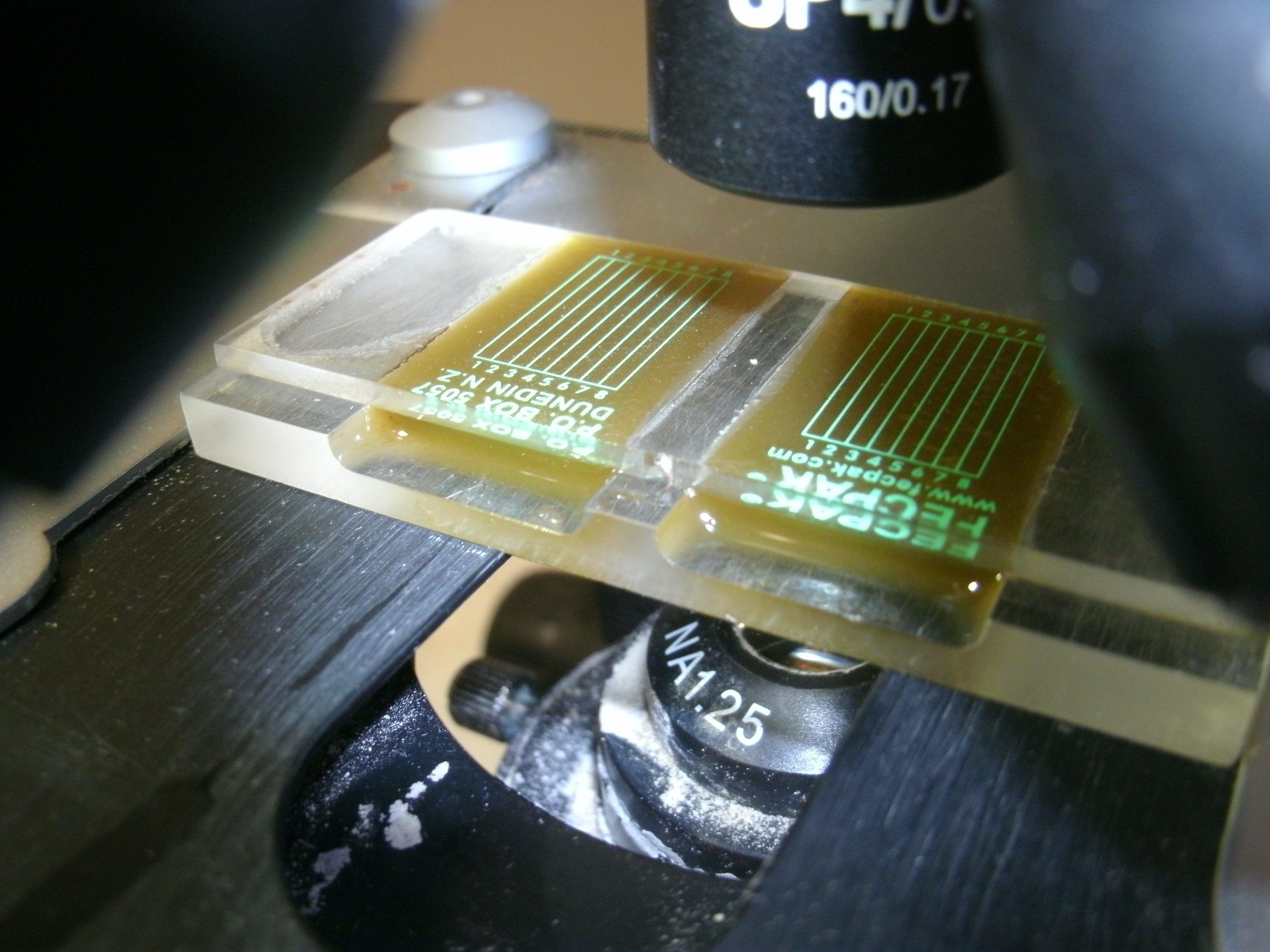
We test the poo by mushing it up with some salty water. The salt in the water makes the eggs float. This is the grossest part of the job!
We then put some of this mix on glass slides, look through the microscope and count all the eggs we see. We can then calculate how bad the parasitic infection is.
This is a lot quicker than other ways of diagnosing animals. And fast information can be the key to the right treatment for saving animals’ lives.
As Tom told me, “We had a guy lose 200 sheep overnight. Vets had taken blood samples, but those results would take days. We did a test and found out it was barber’s pole worm straight away.”
Probing for parasites
There are lots of different animal parasites, but these are the ones that we test for.

Strongyles (STRON-jyels): These are the most common eggs we see. Strongyles live in the abomasum (AB-om-MAY-sum), the fourth stomach of animals. They damage the stomach lining, decreasing the nutrients these animals can extract from their food. Barber’s pole worm is a type of strongyle.
Nematodirus (NEM-ah-tow-DYEruss): These eggs are a lot bigger than strongyles. Nematodirus cause similar types of damage, but live in the small intestine.
Coccidia (cox-SID-ee-ah): These eggs are tiny, but we see lots of them. Generally they don’t affect the host, but if a young animal is under stressful conditions, they can make it really sick.
Tapeworm: “They look nasty, but they don’t cause a lot of the damage,” says Tom. These worms sometimes drop segments of their body, which can be seen in poo. However, we’re on the lookout for triangular eggs in sheep or rectangular eggs in cows. This helps us accurately calculate how bad the infection is.
Liver fluke: “The damage caused by liver fluke can be quite dramatic,” says Tom. “It simply bores holes through the liver once it gets there.” We test for this parasite using coloured dyes instead of salty water, because liver fluke eggs don’t float.
This article was published in Issue 32 of Double Helix magazine (https://www.csiro.au/en/Education/Double-Helix). Copyright for this article is held by CSIRO.
